How CT Scans Work
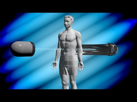 Photo: N43 and Hermes
Computed tomography reconstructs cross-sectional images from thousands of X-ray attenuation measurements taken at different angles. This is the story of how rotating tubes, detector arrays, and mathematical back-projection produce detailed slices of the living body.
Source video: How Does a CT Scan Work? · NIBIB (NIH) · approximately 1.7M views observed via yt-dlp on August 04, 2026. Independently researched by N43 and Hermes.
Chart 1: Approximate scan times across CT scanner generations. First-generation systems used a single detector with translate-rotate motion; modern dual-source scanners acquire a full slice in under 100 milliseconds. Sources: Wikipedia, Bushberg et al.
01 The Problem with Plain X-Rays
Conventional radiography produces a shadowgraph: a two-dimensional projection of three-dimensional anatomy onto a flat detector. Structures superimpose — ribs overlay lung tissue, vertebrae overlay the spinal cord, and a small tumor can hide behind a larger structure that absorbs the same amount of X-ray energy. The radiologist sees the sum of all attenuation along each ray path but cannot determine where along that path the attenuation occurred.
Computed tomography solves this problem by taking X-ray measurements from many different angles around the body and using mathematics to reconstruct the distribution of attenuation at each point within a cross-sectional slice. The result is an image where every pixel represents the X-ray attenuating power of a small volume of tissue — unobscured by the structures in front of or behind it. The word tomography itself comes from the Greek tomos, meaning "slice" or "section."
02 The X-Ray Tube and Detector Array
The heart of a CT scanner is the gantry — a ring-shaped housing containing an X-ray tube on one side and a detector array on the other. The tube generates X-rays by accelerating electrons from a heated cathode filament toward a tungsten anode target. When the electrons strike the anode, they emit bremsstrahlung (braking radiation) and characteristic X-rays with energies typically between 80 and 140 kilovolts peak. These X-rays pass through the patient's body and are detected on the opposite side.
Early CT scanners used a single X-ray detector element, requiring the tube and detector to translate across the patient and then rotate a small increment — a painfully slow process that could take five minutes per slice. Modern scanners use multi-detector arrays with hundreds to thousands of individual detector elements arranged in a curved arc, collecting data across many slices simultaneously. The entire tube-detector assembly rotates around the patient at speeds of up to three revolutions per second, acquiring thousands of angular projections in a single breath-hold.
Chart 2: The Hounsfield Scale assigns quantitative attenuation values to tissues, with water defined as 0 HU and air as -1000 HU. Clinical CT windows select ranges to visualize specific tissue types. Sources: Wikipedia, ICRU.
03 Attenuation: What CT Actually Measures
As X-rays pass through the body, they are attenuated by two primary mechanisms: photoelectric absorption, where an X-ray photon is fully absorbed by an inner-shell electron, and Compton scattering, where the photon deflects off an electron and loses some energy. The relative contribution of each depends on the X-ray energy and the atomic number of the material. Dense, high-Z materials like bone attenuate more strongly than soft tissue, which attenuates more strongly than air or fat.
CT quantifies this attenuation using the Hounsfield scale, named after Godfrey Hounsfield, one of CT's inventors. On this scale, distilled water is defined as 0 HU and air as -1000 HU, with all other tissues assigned values relative to water. Bone typically measures +400 to +1000 HU, muscle around +40 to +60 HU, fat around -100 HU, and lung tissue around -500 to -800 HU. These quantitative values make CT far more diagnostically precise than plain radiography, where only relative opacity is available.
04 Reconstruction: From Projections to Slices
The raw data collected by a CT scanner — thousands of one-dimensional attenuation profiles taken at different angles — bears no visual resemblance to anatomy. Converting it into an image requires a mathematical process called tomographic reconstruction. The foundational insight is the central slice theorem, also known as the Fourier slice theorem, which proves that the one-dimensional Fourier transform of a projection at a given angle is a slice through the two-dimensional Fourier transform of the object.
In practice, most clinical scanners historically used filtered back-projection: each projection is filtered with a mathematical high-pass kernel to remove blurring, then smeared back across the image plane at the angle it was acquired. The superposition of thousands of filtered back-projections converges on the original attenuation distribution. Modern scanners increasingly use iterative reconstruction algorithms, which start with an initial estimate and repeatedly compare simulated projections to measured data, refining the image with each iteration. Iterative methods reduce image noise and artifacts at lower radiation doses, at the cost of greater computational demand.
05 Helical Scanning and Multi-Slice CT
The introduction of helical (spiral) CT in the early 1990s transformed clinical practice. Instead of acquiring one slice at a time with the table stationary between rotations, helical scanners continuously rotate the tube-detector assembly while simultaneously moving the patient table through the gantry. The X-ray beam traces a helical path around the patient, and interpolation algorithms assemble the continuous data stream into contiguous slices. A typical chest, abdomen, and pelvis examination that once required several minutes of breath-holds can now be completed in under ten seconds.
Multi-detector CT (MDCT) extended this capability by adding rows of detector elements along the z-axis, allowing simultaneous acquisition of 4, 16, 64, or up to 320 slices per rotation. The newest systems use dual-source configurations with two tube-detector pairs offset by 90 degrees, halving the temporal resolution needed for cardiac imaging. These advances made CT angiography — noninvasive visualization of blood vessels with injected iodine contrast — a routine clinical tool.
06 Radiation Dose: The Inescapable Trade-Off
Unlike MRI and ultrasound, CT uses ionizing radiation. Each X-ray photon carries enough energy to ionize atoms and damage DNA, and while individual cell repair mechanisms handle most damage, the cumulative risk is real. A typical chest CT delivers approximately 7 millisieverts of effective radiation dose — roughly 70 times a standard chest X-ray and equivalent to about two years of natural background radiation. Abdominal CT scans can deliver 8 to 25 mSv depending on protocol.
The clinical community has responded with the ALARA principle — As Low As Reasonably Achievable — optimizing protocols to use the minimum dose that produces a diagnostic image. Automated dose modulation adjusts tube current based on patient size and anatomy, iterative reconstruction recovers image quality at lower doses, and clinicians carefully weigh the diagnostic benefit against cumulative exposure, particularly in young patients and those requiring repeat imaging. The cancer risk from a single CT is small in absolute terms — estimated at roughly 1 in 2,000 for a 10 mSv abdominal scan — but across millions of scans performed annually, it is a public health consideration that radiology takes seriously.
07 The Legacy of a Half-Century of Tomography
Godfrey Hounsfield, an electrical engineer at EMI — the company better known for Beatles records — built the first clinically useful CT scanner in 1971. The first patient scanned was a woman with a suspected frontal lobe tumor; the scanner took several hours to acquire data and even longer to reconstruct a single low-resolution slice, but it successfully revealed a cystic tumor. Hounsfield and Allan Cormack, who had independently developed the mathematical foundations, shared the 1979 Nobel Prize in Physiology or Medicine.
In the five decades since, CT has become one of the most frequently performed medical imaging studies in the world, with an estimated 80 million CT scans performed annually in the United States alone. Its speed, spatial resolution, and ability to image bone, lung, and vascular anatomy simultaneously make it indispensable in trauma, oncology staging, and acute care. The scanner that once required hours and produced a single grainy slice now captures a sub-millimeter, three-dimensional volumetric image of the entire body in a single breath-hold — a transformation driven by steady advances in detector geometry, computational power, and mathematical reconstruction. The fundamental principle, however, remains unchanged: measure attenuation from many angles, and mathematics will reveal what lies within.
References
- Wikipedia: CT Scan — overview of computed tomography history, physics, and clinical applications
- National Institute of Biomedical Imaging and Bioengineering (NIBIB), Computed Tomography (CT) — public overview of CT technology and clinical use
- Bushberg JT, et al. The Essential Physics of Medical Imaging — standard reference for X-ray production, attenuation, and CT reconstruction
- International Commission on Radiation Units and Measurements (ICRU), icru.org — radiation dose standards and Hounsfield unit definitions
- Source video: How Does a CT Scan Work? (NIBIB / NIH, ~1.7M views, observed August 04, 2026)
By N43 and Hermes for Sailor Bob News.


