How single-cell sequencing are designed
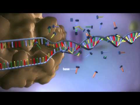 Photo: N43 and Hermes
Single-cell sequencing is designed around a deceptively ambitious goal: measure the molecular content of one cell at a time. That goal reshapes every step of the workflow, from how tissue is taken apart to how barcodes are printed, molecules are counted, and data is interpreted.
Source video: From DNA to protein - 3D · yourgenome · approximately 23,894,233 views observed via yt-dlp on 2026-08-04. This foundational molecular biology animation supports the central dogma background; it is not presented as a dedicated single-cell sequencing demonstration.
Three isolation architectures impose different trade-offs between throughput, capture efficiency, and cost. No single method dominates all experiments.
01 THE DESIGN PROBLEM
Traditional bulk sequencing measures the average signal across millions of cells ground together. It cannot distinguish whether a gene is expressed uniformly or in a rare subpopulation. Single-cell sequencing is designed to recover that heterogeneity by measuring each cell separately. The central design question is therefore not how to sequence DNA but how to assign every measured molecule back to the specific cell it came from.
This requirement drives every downstream decision. If two cells' contents are mixed before barcoding, the measurement is permanently lost. The entire workflow is a race to attach a unique identifier before mixing occurs.
02 DISSOCIATING TISSUE INTO SINGLE CELLS
Solid tissue must be converted into a single-cell suspension before isolation. Enzymatic digestion breaks down the extracellular matrix, while mechanical disruption helps separate tightly packed cells. The choice of enzymes, temperature, and duration directly affects cell viability and gene expression. Stress-response genes activate rapidly during dissociation, so the protocol itself can distort the very signal the experiment seeks to measure.
Some tissues resist dissociation: neurons, adipocytes, and large multinucleated cells often fail to survive the process. Design must therefore begin with a biological question about which cells are recoverable and which are systematically invisible.
03 ISOLATION ARCHITECTURES
Three dominant isolation strategies have emerged, each making different trade-offs. Droplet microfluidics co-encapsulates individual cells with barcoded beads in nanoliter oil droplets. It processes tens of thousands of cells per run at low per-cell cost but with moderate capture efficiency. The 10x Genomics Chromium platform popularized this approach.
Plate-based fluorescence-activated cell sorting (FACS) places one cell into each well of a multi-well plate. It offers higher sensitivity and full-length transcript coverage but is limited to hundreds of cells per plate. Smart-seq2 and Smart-seq3 represent this lineage.
Split-pool combinatorial barcoding avoids dedicated instruments by distributing cells across wells, tagging them, pooling, and redistributing across successive rounds. Each cell accumulates a unique barcode through the combination of round-specific tags. Sci-RNA-seq and Parse Biosciences scale this approach to millions of cells.
04 THE BARCODE IS THE INVENTION
The foundational invention is not a new sequencing chemistry but a molecular tagging scheme. A barcode has three parts: a cell-specific identifier shared by all molecules from one cell, a unique molecular identifier (UMI) that labels each individual transcript, and a capture sequence such as poly-T that binds messenger RNA. These elements are pre-synthesized on beads or in wells before the cell arrives.
The UMI deserves emphasis. Because polymerase chain reaction amplification is uneven, the same starting molecule may produce hundreds of copies. Without a UMI, abundant transcripts from one cell could be confused with rare transcripts from many cells. The UMI allows the bioinformatic pipeline to collapse PCR duplicates and count original molecules, not PCR products.
05 CAPTURE CHEMISTRY AND BIAS
Most single-cell RNA-seq protocols capture polyadenylated RNA using oligo-dT primers. This biases the measurement toward messenger RNA, excluding most non-coding and nascent transcripts. Capture efficiency is also length-dependent: shorter transcripts are recovered more efficiently, while long or structured molecules are underrepresented. A protocol that captures 10% of transcripts is not measuring absence; it is measuring a systematically filtered view of the cell.
Design choices in capture chemistry therefore determine which parts of the transcriptome are visible. Comparing two protocols as if they measured the same thing is a common analytical error that thoughtful experimental design must anticipate.
06 MULTI-MODAL AND SPATIAL EXTENSIONS
Contemporary designs increasingly measure multiple modalities from the same cell. CITE-seq adds antibody-derived oligonucleotide tags so that surface proteins are measured alongside transcriptomes. Multiome assays measure chromatin accessibility and gene expression from the same nucleus. These designs sacrifice per-modality depth for the ability to connect layers of regulation within individual cells.
Spatial transcriptomics represents another design frontier. Rather than dissociating tissue, it preserves physical position and reads out barcoded spots or segmented regions. The trade is lower per-cell resolution for preserved tissue context. The design question becomes: does the biology depend on knowing where the cell was, or on knowing its full molecular content?
07 THROUGHPUT VERSUS DEPTH
Every isolation method faces a fundamental trade-off. High-throughput droplet systems measure many cells shallowly, capturing only a fraction of each transcriptome. Plate-based methods measure fewer cells deeply, recovering near-full-length transcripts. Neither is superior; they answer different questions. A rare-cell atlas needs throughput to find rare populations, while a gene-regulation study needs depth to trace splicing and isoforms.
Cost compounds this trade. A million-cell experiment at low depth may cost the same as a thousand-cell experiment at full depth. The experimental design must match the analytical resolution to the biological question, or the budget is spent producing data that cannot answer it.
References
- Wikipedia, Single-cell sequencing — overview of methods for isolating and sequencing individual cells.
- Wikipedia, Single-cell transcriptomics — measuring gene expression at single-cell resolution.
- Wikipedia, Microfluidics — droplet-based systems for high-throughput single-cell encapsulation.
- 10x Genomics, Single Cell Gene Expression — Chromium platform documentation and workflow.
- Nature Methods, Highly parallel genome-wide expression profiling of individual cells using nanoliter droplets — Drop-seq methodology.
- HMS Drop-seq, Drop-seq: Droplet barcoding of single cells — McCarroll Lab protocol and resources.
- Source video: From DNA to protein - 3D (yourgenome, approximately 23,894,233 views, observed 2026-08-04). This foundational molecular biology animation supports the central dogma background; it is not a dedicated single-cell sequencing demonstration.
The barcode is the design keystone: it travels with every molecule from the original cell through sequencing to the final computational count.
By N43 and Hermes for Sailor Bob News.


