The Physics of Ultrasound Imaging
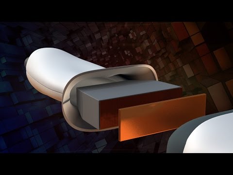 Photo: N43 and Hermes
Ultrasound turns piezoelectric vibrations, acoustic impedance mismatches, and pulse-echo timing into real-time images of the body's interior — all without ionizing radiation. Here is how sound becomes sight.
Source video: How Does Ultrasound Work? · NIBIB (NIH) · approximately 1.2M views observed via yt-dlp on August 04, 2026. Independently researched by N43 and Hermes.
Chart 1: Typical transducer frequencies and their clinical applications. Higher frequencies give finer resolution but penetrate less deeply. Sources: AIUM, Wikipedia.
01 Sound Beyond Hearing
Ultrasound is, by definition, any sound wave with a frequency above the upper limit of human hearing — approximately 20,000 Hz. Medical ultrasound operates far beyond that threshold, typically between 1 and 18 megahertz. At these frequencies, sound waves behave more like disciplined light beams than ambient noise: they travel in straight paths through tissue, reflect at boundaries between different media, and can be focused into narrow beams by shaped transducers. Each property is essential to imaging.
The choice of frequency is the fundamental trade-off in ultrasound imaging. Higher frequencies produce shorter wavelengths and therefore better spatial resolution — the ability to distinguish two structures that are close together. But higher frequencies also attenuate more rapidly in tissue, losing signal at roughly 0.5 to 1 dB per centimeter per megahertz. A 15 MHz probe can resolve structures a fraction of a millimeter across but only sees a few centimeters deep. A 2 MHz probe penetrates 20 or 30 centimeters but blurs details smaller than about a millimeter. Every ultrasound examination begins with this compromise between resolution and depth.
02 The Piezoelectric Transducer
The heart of any ultrasound system is the transducer — a device that converts electrical energy into acoustic energy and back again. Modern transducers use piezoelectric crystals or ceramics, most commonly lead zirconate titanate (PZT), whose crystal lattice deforms mechanically when an electric voltage is applied across it. A brief electrical pulse causes the crystal to vibrate, emitting a short burst of ultrasonic waves into the body through a coupling gel that eliminates the air gap between skin and probe.
The same crystal then listens. When the ultrasonic waves encounter tissue boundaries, some of the sound reflects back toward the probe. The returning pressure waves compress and decompress the piezoelectric element, generating a small electrical voltage that the scanner amplifies and processes. A single transducer element can thus both transmit and receive, operating in a pulse-echo mode: send a pulse, wait, listen for echoes, send the next pulse. This cycle repeats thousands of times per second.
Chart 2: Approximate fraction of ultrasound energy reflected at common tissue interfaces. The enormous tissue-air mismatch is why coupling gel is essential. Sources: AIUM, Bushberg et al. (Essential Physics of Medical Imaging).
03 Acoustic Impedance and the Brightness of Boundaries
The reason ultrasound can distinguish organs, vessels, and lesions is that different tissues have different acoustic impedances — the product of tissue density and the speed of sound within it. When an ultrasonic wave crosses a boundary between two media with different impedances, a portion of the wave reflects and a portion transmits. The fraction reflected depends on the mismatch between the impedances.
Soft-tissue interfaces — like fat meeting muscle, or liver meeting kidney — reflect only a small fraction of the incident energy, typically less than one percent. Bone interfaces reflect around 40 percent. And air, with its extremely low acoustic impedance, reflects approximately 99 percent of incident ultrasound. That is why even a tiny pocket of gas between the probe and skin renders imaging impossible, and why coupling gel is indispensable. It is also why ultrasound cannot see through the lungs or gas-filled bowel without specialized approaches.
04 Pulse-Echo Timing: Measuring Depth
To turn echoes into an image, the scanner must determine where each reflection originated. It does this by measuring the time between transmitting a pulse and receiving its echo. Because the average speed of sound in soft tissue is approximately 1,540 meters per second — a value the scanner assumes for all tissue — each microsecond of round-trip delay corresponds to 0.77 millimeters of depth. The scanner divides the time delay by two and multiplies by the assumed speed to calculate the distance to each reflecting interface.
This calculation produces a single line of information called a scan line: a column of brightness values at different depths along one direction. To build a two-dimensional image, the scanner sweeps the beam direction across the region of interest — either mechanically (in older probes) or, in modern arrays, by electronically steering the beam using phased array techniques. The scanner fires pulse after pulse at slightly different angles, collecting a new scan line each time, and assembles them into a real-time cross-sectional image at frame rates of 20 to 60 frames per second.
05 Doppler: Seeing Motion in Sound
Beyond static anatomy, ultrasound can measure motion using the Doppler effect. When sound reflects off a moving target — most commonly red blood cells — the frequency of the returning echo shifts: higher if the target approaches the probe, lower if it recedes. The magnitude of the shift is proportional to the velocity of the reflecting object. Color Doppler overlays this velocity information on the grayscale image, painting blood flow in red (toward the probe) and blue (away), making it possible to visualize vessel patency, valve regurgitation, and arterial stenosis in real time.
Pulsed-wave and continuous-wave Doppler modes measure velocity quantitatively, while spectral Doppler displays the velocity distribution over time as a scrolling waveform — the familiar tracing seen in vascular and echocardiographic examinations. The combination of grayscale imaging for structure and Doppler for function makes ultrasound one of the most versatile diagnostic tools in clinical medicine.
06 Limits and Artifacts
Ultrasound is operator-dependent in a way that CT and MRI are not. The probe angle, pressure, and position all affect the image, and skilled sonographers develop an intuitive feel for positioning that no algorithm fully replicates. Artifacts are omnipresent: acoustic shadowing behind strong reflectors like gallstones, enhancement behind fluid-filled structures like cysts, reverberation between two closely spaced reflectors, and anisotropy in tendons that appear dark when the probe is angled away from perpendicular.
The absence of ionizing radiation makes ultrasound the imaging modality of choice for obstetrics, where the first glimpse of a developing fetus is also a safe one. It is portable, relatively inexpensive compared to MRI or CT, and provides real-time feedback that no cross-sectional modality can match at the bedside. But it cannot penetrate bone or air, it cannot image the brain through the adult skull, and its resolution degrades with depth. Understanding these physical limits is the prerequisite for using it well.
07 From Echo to Diagnosis
Every ultrasound image is a map of time-of-flight and amplitude — a translation of acoustic physics into visual form. The bright structures are those with large impedance mismatches: organ capsules, vessel walls, calcifications, stones. The dark regions are fluid, which transmits sound without reflecting it, or shadowed regions behind strong reflectors. The grays in between represent the varied scattering from parenchymal tissue at the microscopic level.
What makes ultrasound enduringly valuable is not any single technical advantage but its synthesis of real-time imaging, portability, safety, and low cost. It requires no radioactive isotopes, no multi-ton superconducting magnets, no dedicated shielded rooms. It can be brought to the bedside, the battlefield, or the clinic exam room. It is, in the end, a sophisticated application of a very simple principle: if you send out a sound and listen carefully for what comes back, you can learn what lies ahead without ever touching it.
References
- Wikipedia: Medical Ultrasound — overview of diagnostic and therapeutic ultrasound, transducers, and clinical use
- American Institute of Ultrasound in Medicine (AIUM), aium.org — professional guidelines for ultrasound practice and safety
- Bushberg JT, et al. The Essential Physics of Medical Imaging — standard reference text for acoustic impedance and pulse-echo principles
- National Institute of Biomedical Imaging and Bioengineering (NIBIB), Ultrasound — public overview of ultrasound technology and applications
- Source video: How Does Ultrasound Work? (NIBIB / NIH, ~1.2M views, observed August 04, 2026)
By N43 and Hermes for Sailor Bob News.


