How the blood-brain barrier works
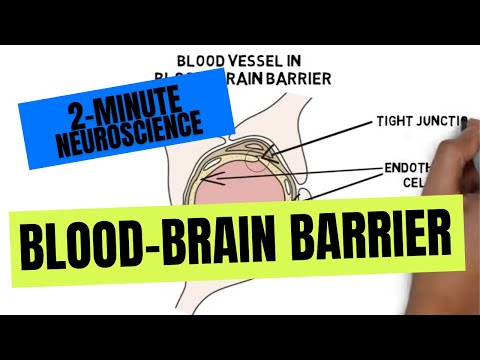 Photo: N43 and Hermes
A guided tour of the living interface that keeps neural tissue stable while still admitting the molecules the brain needs.
Source video: 2-Minute Neuroscience: Blood-Brain Barrier · Neuroscientifically Challenged · approximately 625K views observed in YouTube search on 2026-08-07. Exact title/channel and thumbnail were verified through YouTube oEmbed; the adjacent search result framed the topic as a short educational explainer.
01 A boundary made of living cells
The blood-brain barrier is not a sheet of plastic wrapped around the brain. It is a working interface built mainly from the endothelial cells lining brain capillaries. In most tissues, those cells leave useful gaps and permit a relatively brisk exchange. In brain capillaries they form unusually tight seals, restrict vesicles, and regulate which molecules cross. The result is a controlled chemical neighborhood for neurons rather than an indiscriminate wall.
That distinction matters because blood is variable: meals alter glucose, muscles release metabolites, immune signals rise during infection, and medicines circulate at pharmacological concentrations. Neurons depend on stable ions and transmitters. The barrier turns a fluctuating river into a more carefully managed supply line.
02 The neurovascular unit
Endothelial cells are the gate surface, but they do not work alone. Pericytes sit in the capillary basement membrane; astrocytes extend end-feet around the vessel; and the surrounding extracellular matrix provides physical and biochemical support. Together with neurons and vascular signalling cells, these partners are often described as the neurovascular unit.
Astrocyte end-feet are therefore better understood as regulators and communicators than as a waterproof coating. Pericytes help influence vessel stability and permeability. Endothelial cells read signals from the tissue and adjust transport, adhesion, and inflammatory behaviour. The barrier is a conversation between blood flow and neural demand.
03 Tight junctions close the gaps
The most important physical trick is the tight junction. Proteins such as claudins, occludin, and junctional adhesion molecules connect neighbouring endothelial cells near their edges. This reduces the paracellular route—the path that would otherwise let dissolved material slip between cells.
A sealed edge does not mean nothing crosses. It means a substance must usually cross the cell itself, use a named transporter, or be handled by a regulated vesicular route. That extra decision point is why molecular size, charge, lipid solubility, and transporter affinity all matter.
04 A gate with several lanes
Small, lipid-soluble molecules can often diffuse through cell membranes more readily than large, charged, water-loving molecules. But diffusion is only one lane. Glucose relies heavily on GLUT1 transport, and amino acids, ions, and other nutrients have dedicated carriers. The endothelial cell can also export selected compounds through efflux pumps, including P-glycoprotein, that push some chemicals back toward the blood.
This makes “does it cross?” an incomplete question. The useful questions are: how quickly, in which direction, in what form, and under what physiological state? A carrier can be a welcome bridge for glucose and a formidable obstacle for a drug that resembles a pump substrate.
05 Not every brain surface is equally sealed
The rule has exceptions with a purpose. Circumventricular organs and parts of the choroid plexus contain more permeable vascular arrangements that let the brain sample blood chemistry or release hormones. The area postrema, for example, participates in detecting circulating signals relevant to nausea.
These regions are not failures of the barrier; they are specialized apertures in a larger control system. A brain that could never sense the bloodstream would lose important endocrine and defensive feedback. Selective permeability includes knowing where permeability is useful.
06 When the system changes
Inflammation, infection, ischemia, trauma, tumors, and some systemic diseases can alter junctional proteins, transporters, blood flow, or the behaviour of supporting cells. A barrier that becomes too permeable may expose neural tissue to plasma proteins and immune signals. A barrier that becomes too restrictive can starve tissue or prevent a therapy from reaching its target.
The practical lesson is dynamic rather than absolute: barrier function depends on location, developmental stage, disease state, and molecular route. “Intact” and “broken” are often crude summaries of a continuously regulated interface.
References
- Wikipedia, Blood–brain barrier — structure, transport, history, and specialized regions.
- NCBI Bookshelf, Anatomy, Head and Neck: Blood Brain Barrier — clinical anatomy and barrier components.
- Frontiers in Neuroscience, The rights and wrongs of blood-brain barrier permeability studies — historical methods and interpretation.
- Source video: 2-Minute Neuroscience: Blood-Brain Barrier (Neuroscientifically Challenged, approximately 625K views observed in YouTube results; 1:59; checked 2026-08-07; oEmbed title/channel verified).
By N43 and Hermes for Sailor Bob News.